

Radiographs are an essential component of dental diagnosis and treatment planning for a general dentist in Montville, NJ. You may be given radiographs as part of your clinical assessment. Periapical radiography is a frequent intraoral imaging method in radiology that may be included in your radiologic exam. In this article, we will talk about the uses and processes of periapical X-rays.
Uses:
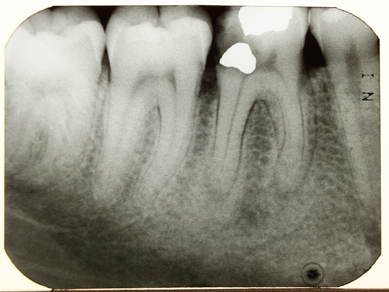
The primary application is to diagnose diseases affecting teeth and adjacent tissues. Sometimes dentists are unsure whether there is a periapical process, thus they must utilize this X-ray to determine a diagnosis. They also utilize it as part of the therapy process, such as when they need to examine the depth of dental cavities. When it comes to decay, dentists can see which tissues it has impacted, which surfaces, and whether it is close to the tooth pulp.
This is all extremely beneficial for eliminating the rot and applying the filler. In periapical situations, an X-ray can determine if the infection has migrated from the pulp to the surrounding bone, whether it is a localized infection, or spread. It aids in the diagnosis of dental and bone fractures, cysts, tumors, the number of roots, canals, apex position, and other issues.
What Is the Process Like?
Periapical X-rays are a form of intraoral X-ray. This indicates that the X-ray film is placed into the mouth. Here’s what to expect from a normal periapical radiograph:
Before the X-ray, the technician will provide you with a lead apron and thyroid collar. These are intended to reduce your exposure to X-ray radiation. The dental assistant will insert a piece of film into an XCP (extension cone paralleling kit). The section containing the film will have a perpendicular bite plate. The film and holder are put adjacent to the tongue, and the patient bites down on the plate to keep the film stable and in the proper position.
Once the XCP kit is in place and you are firmly biting down on the bite plate, the technician will leave the room and activate the X-ray equipment. They will return immediately to remove the film and XCP equipment from your mouth. After reviewing the radiological pictures, the dentist will make a diagnosis and prescribe a treatment plan.
The best thing is that these types of x-rays target fewer teeth, allowing for a sharper image of specific teeth, and making diagnosis simpler and easier for the dentist.


Comments